
Figure 2.
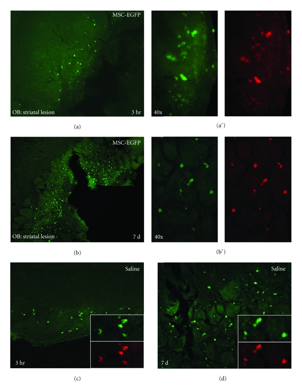
Autofluorescence in the olfactory bulb (OB) after intranasal delivery of MSC-EGFPs to mice with prior mechanical lesions in striatum. Fluorescent structures are observed within the glomerular layer of the OB at 3 hours and 7 days after intranasal administration of MSC-EGFPs (a), (b) or saline (c), (d). Higher magnification images of the OB at 3 hours ((a′, c) inset) and 7 days ((b′, d) inset) imaged with both the GFP and Texas Red bandpass filters show a one-to-one correlation between the number and intensity of stained structures.