
Figure 1.
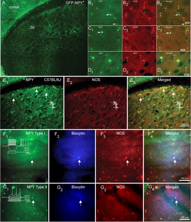
Distribution and expression of NPY/SOM/NOS+ in striatal NPY interneurons in mice. A, Fluorescent photomicrographic montage of a coronal section of a BAC NPY-GFP+ mouse brain illustrating the distribution and abundance of NPY interneurons in the neostriatum (Str). Note the sparse distribution of striatal NPY interneurons compared with cortical NPY interneurons. B1–B3, Higher-magnification photomicrographs illustrate GFP–NPY fluorescence in B1, NOS immunofluorescence in B2, and GFP and NOS merged in B3. The small arrows indicate dimly fluorescent Type I somata, and the larger arrows indicate brighter Type II somata. Note that only Type I interneurons express NOS. C1–C3, GFP–NPY fluorescence in C1, SOM immunofluorescence in C2, and GFP and SOM merged in C3. Note that only Type I interneurons express SOM. D1–D3, GFP–NPY fluorescence in D1, NPY immunofluorescence in D2, and GFP and SOM merged in D3. Note that both type I and type II interneurons express NPY. E1–E3, NPY and NOS immunofluorescence in striatum from normal C57BL/6J mice shows exactly that same pattern of colocalization as in BAC NPY-GFP+ mice. E1, NPY immunofluorescence reveals two different types of NPY somata, a few very bright ones (presumed Type II NPY interneuron), marked by the white arrows, and more numerous less bright ones (presumed type I NPY interneuron), some marked by white double arrows. E2, NOS immunofluorescence. Three NOS-immunoreactive neurons are marked by white double arrows. E3, Overlay of E1 and E2 shows that the bright NPY neurons do not colocalize NPY (white arrows), whereas the less bright cells do colocalize NOS (white double arrows). F, Example of an electrophysiologically identified Type I NPY interneuron (arrow) identified by GFP fluorescence (F1) filled with biocytin (F2) that also is immunoreactive for NOS (F3), with the triple overlay shown in F4. G, Example of an electrophysiologically identified type II NPY interneuron (arrow) identified by GFP fluorescence (G1) filled with biocytin (G2) that is not immunoreactive for NOS (G3), with the triple overlay shown in G4.