
Figure 3.
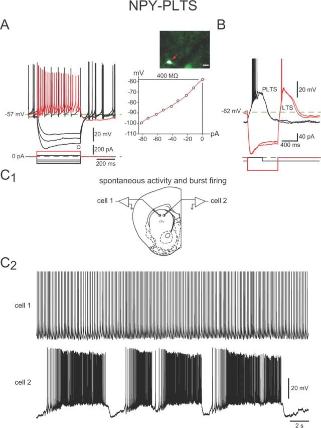
Intrinsic electrophysiological properties of NPY–PLTS interneurons. A, Responses to negative and positive current pulses in an NPY–PLTS interneuron (inset) reveal high input resistance, a time-dependent Ih-like sag in response to hyperpolarizing current injections, and a relatively depolarized resting membrane potential. Spike train elicited by depolarization (red trace) exhibits modest spike-frequency adaptation and marked accommodation. These interneurons exhibited spontaneous activity with little rebound after offset of hyperpolarizing current injections (black traces). B, Slightly more hyperpolarized NPY–PLTS interneuron exhibits a prolonged plateau potential (PLTS) following a depolarizing current injection (black trace) and rebound LTS and plateau following offset of hyperpolarizing current injections (red traces). C1, Approximate locations of the two cells shown in C2. C2, Two NPY–PLTS interneurons recorded simultaneously exhibit spontaneous activity. Top, Typical tonic firing pattern. Bottom, In a few cases, NPY–PLTS interneurons showed 8–10 mV membrane potential oscillations that separated prolonged epochs of higher frequency spontaneous burst firing activity.