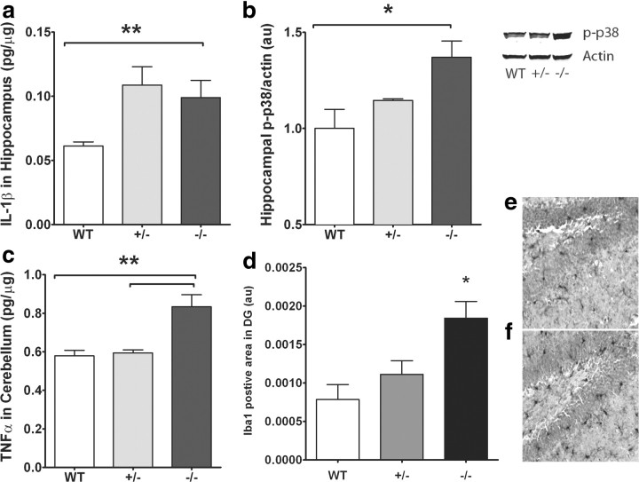
Figure 5.
a, CX3CR1−/− and CX3CR1+/− mice show increased hippocampal protein levels of IL-1β compared to littermate WTs as measured by ELISA. b, Western blot analysis of hippocampi from wild-type, CX3CR1−/−, and CX3CR1+/− mice shows an increase in phospho (p)-p38 protein in CX3CR1−/− and CX3CR1+/− mice compared to that of wild-type (right blot; near-infrared image displayed in gray scale). Relative p-p38 expression normalized to β-actin shows the highest expression in CX3CR1−/− and CX3CR1+/− mice. c, TNFα cerebellar protein levels are significantly increased in CX3CR1−/− and CX3CR1+/− mice compared to wild-type mice as measured by ELISA. d, The area of Iba-1 staining is significant higher in CX3CR1−/− mice compared to wild-type and CX3CR1+/− mice as measured by immunohistochemistry. White bar, WT; gray bar, CX3CR1+/−; black bar, CX3CR1−/−. e, f, Representative photomicrographs of the Iba-1+ cells in the dentate gyrus of wild-type (e) and CX3CR1-deficient mice (f). All data are presented as mean ± SEM. p < 0.01. **p < 0.005; *p < 0.05.

