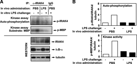
Figure 4. Suppressed LPS-inducible activation of IRAK4 in macrophages obtained from endotoxin-treated mice.
(A) Twenty-six hours after i.p. inoculation of PBS or LPS (25 μg/mouse), spleen were isolated and homogenized, and spleen cells were adhered to plastic to obtain macrophages, followed by stimulation of cells for 15 min with medium or 100 ng/ml LPS. IRAK4 proteins were immunoprecipitated (IP) from cell lysates and subjected to in vitro kinase assays with MBP as a substrate. Cell lysates were analyzed by immunoblotting, using anti-IκBα, anti-IRAK4, and antitubulin antibodies. The results of representative (n=3) experiments are presented. (B) Densitometric quantification of the data shown in A. Intensities of p-IRAK4 and p-MBP bands were measured and normalized to the levels of total IRAK4 and tubulin. Data in the treatment groups were divided by values obtained in medium-treated macrophages obtained from PBS-injected mice and presented as fold induction.

