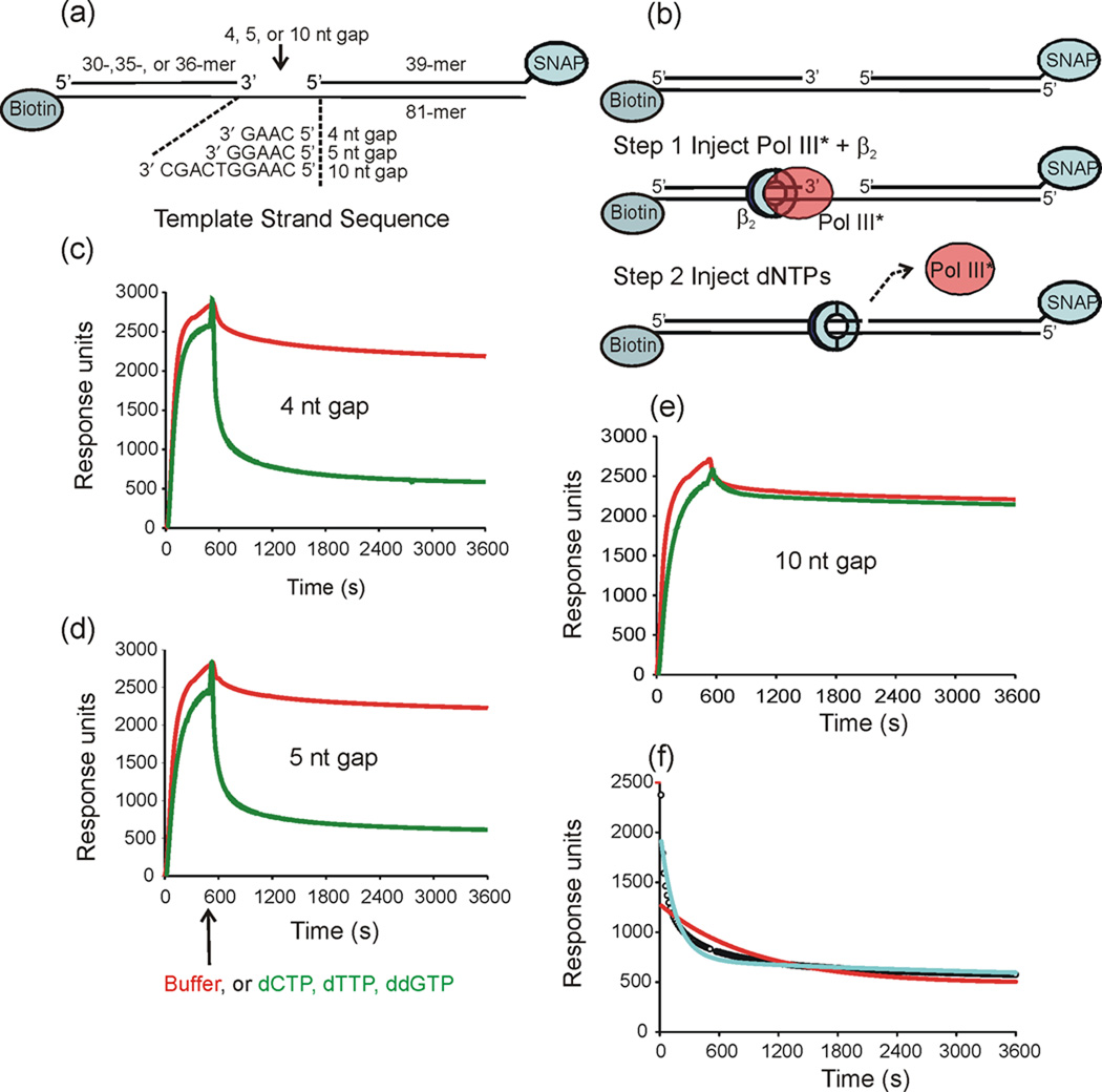
Fig. 1.
Pol III* does not release rapidly upon filling a gap. The dissociation of Pol III* from primed templates was measured using surface plasmon resonance. (a) Three primed templates containing 4-, 5- and 10-nt gaps were anchored to the surface of a streptavidin-coated sensor chip using biotin-labeled oligonucleotides. The 3′-end of the blocking oligonucleotide was covalently linked to the SNAP protein to prevent β2 from sliding off the end. (b) Initiation complexes were assembled onto the immobilized DNA primer-template by injecting Pol III*, β2 and ATP. A second injection contained either buffer or a solution containing 40µM dCTP, dTTP, and ddGTP (dNTPs) was made at 540 s. (c) (d) and (e) Injection of either buffer (red) or dNTPs (green) over the 4-nt gapped template 5-nt gapped template, and 10-nt gapped template, respectively. Dissociation was allowed to continue for three days. The data shown is only for the first h after injection. (f) Curve-fit analysis is shown for the for Pol III* dissociation in the presence of dNTPs over a 4-nt gapped template: data (open circles), curve fit to single exponential decay (red), curve fit to double exponential decay (cyan).

