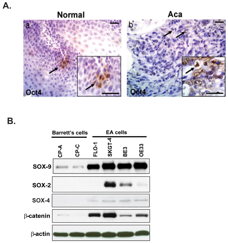
Figure 3.
Evaluation of Oct4 expression in normal and esophageal adenocarcinoma (ACa). (A) Oct4+ was detected in tissues of normal (a) and esophageal adenocarcinoma (Aca) tissues (b) by immunohistochemistry as described in material and methods. Inset shows the respective figures at higher magnification. Scale bar is 50 μM. (B). Immunoblots were performed to analyze SOX-9, SOX-2 and SOX-4 and β-catenin expression using cell lysate were from Barrett's cells (CP-A, CP-C) and adenocarcinoma cells (FLO-1, SKGT-4, BE3 and OE33) as described in materials and methods.

