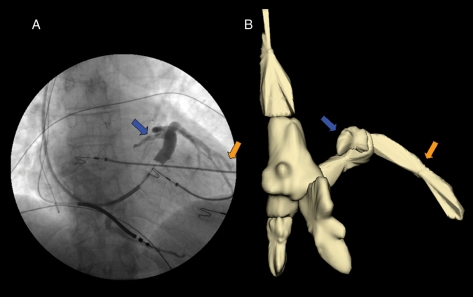
Figure 1.
Case 4. (A) The coronary sinus angiography performed with the Swan-Ganz catheter in a antero-posterior projection. The blue and orange arrows indicate the coronary sinus and the presence of a lateral vein, respectively. This image also shows the presence of a bipolar implantable cardioverter–defibrillator lead placed in the apex of the right ventricle and it is possible to see the tip of a J-wire in the right atrium. (B) Reconstruction of the right cardiac chambers, the superior and inferior vena cava and the coronary sinus using the NavX system; the blue and orange arrows indicate the three-dimensional reconstruction of the coronary sinus and the lateral vein, respectively.

