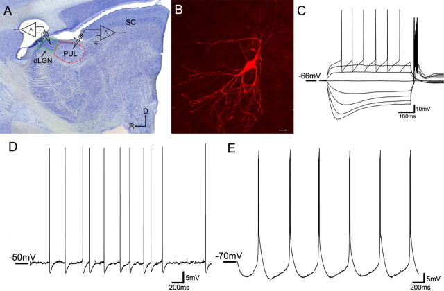
Figure 1.
In vitro recording methods. A, A parasagittal section of the tree shrew brain stained for Nissl substance illustrates the location of the whole-cell recordings in either the dLGN (outlined in green) or the pulvinar nucleus (PUL; outlined in red). D, dorsal, R, rostral, SC, superior colliculus. B, A confocal image of an adult pulvinar cell filled with biocytin during recording (scale bar, 20 μm). All successfully filled cells exhibited morphologies consistent with their identification as relay cells. C–E, Voltage fluctuations were recorded in response to the injection of depolarizing or hyperpolarizing current steps of varying size (C, adult pulvinar cell) as well in response to depolarization (D, adult pulvinar cell) or hyperpolarization (E, adult pulvinar cell) of the membrane potential through constant current injection.

