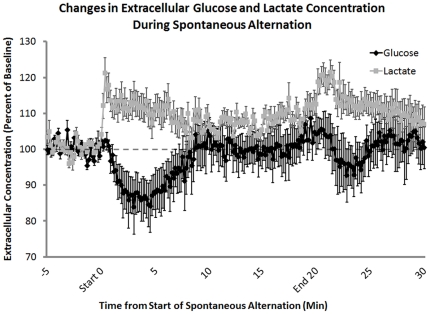
Figure 3. Extracellular lactate and glucose levels in the hippocampus, measured before, during, and after behavioral testing.
Using lactate- and glucose-specific biosensors, extracellular concentrations of both lactate and glucose were measured during spontaneous alternation testing. Lactate concentrations significantly increased at the beginning of behavioral testing (n = 4; t3 = 4.77, p<0.02; MEAN ± SEM: 112.50%±3.15%). In contrast, glucose concentrations decreased after 5 minutes on the task (n = 3; t2 = 3.58, p<0.05; MEAN ± SEM: 86.19%±7.73%). The increase in extracellular glucose seen 5–10 min after the start of memory testing corresponds to an increase in blood glucose levels (baseline vs. 10 min: p = 0.47, 10 min MEAN ± SEM: 103.68%±6.29%). After the rat was removed from the maze there was a significant increase in lactate compared to baseline levels (t3 = 4.77, p<0.02; MEAN ± SEM: 117.9%±2.87%) most likely due to handling.

