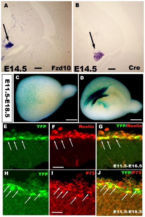
Figure 1. Cortical hem-derived CR cells originating from progenitors expressing Fzd10.
A–B: RNA in situ hybridization showing that Cre expression mimics endogenous Fzd10 in the cortical hem in the Fzd10-CreER™ mouse. C–D: Whole-mount X-gal staining of E18.5 Fzd10 CreER™/R26R-LacZ brains when TM was administered at E11.5, showing the output of hem-derived CR cells. C: Lateral view of the hemisphere. D: Middle view. E–J: YFP reporter-positive cells are both reelin+ and P73+, indicating that these cells are CR cells originating from the cortical hem. E–G: When TM was injected at E11.5 and double immunostaining was performed at E16.5, YFP+ cells expressed reelin. H–J: YFP+ cells were also P73 positive. E11.5–E18.5, TM was administered at E11.5 and brains were stained at E18.5. Scale bars: A, B: 100 µm; C–D: 2 mm; others: 200 µm.

