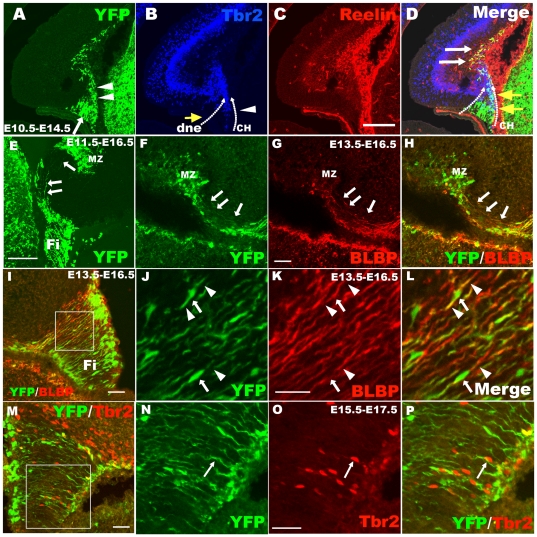
Figure 5. The hem-derived CR cells migrate to the hippocampal marginal zone along the fimbrial radial glial scaffold.
A–D: Triple immunostaining for YFP/reelin/Tbr2 in E14.5 brains when TM was injected at E10.5. A: Cortical hem-derived YFP-positive cells consisting of two types of cells, radial glial scaffold (arrow) and migrating cells (arrowheads). B: Anti-Tbr2 staining shows two Tbr2+ migrating streams, one from the dentate neuroepithelium to produce dentate granule cells indicated by yellow arrows, the other from the cortical hem (arrowheads). They meet at the area of the future dentate. C: Hem-derived reelin-positive CR cells. D: Yellow arrows indicate double-labeled YFP+/Tbr2+ cells migrating out of the cortical hem, and white arrows indicate YFP+/reelin+ cells settled at the MZ zone of the hippocampus. E: Some scaffold-like YFP-positive cells are derived from the cortical hem and distribute to the dentate gyrus from the hem (arrows). F–L: The YFP+ scaffold is positive for the radial glial marker BLBP. F–H: YFP+/BLBP+ radial glial scaffold extend to the DG from the fimbria (arrows). I–L: YFP+/BLBP+ radial glial scaffold in the fimbria. Arrowheads indicate the processes of the radial glia, and the arrow shows its cell body. J–L show high-magnification views of the area boxed in I. M–P: CR cell progenitors, Tbr2+ cells migrate out of the hem along the YFG-positive radial glial scaffold also derived from the cortical hem. Arrows in N–P show migrating Tbr2+ CR progenitors. N–P: High-magnification view of the boxed area in M. dnp, dentate neuroepithelium; CH, cortical hem; Fi, fimbria; MZ, marginal zone; DG, dentate gyrus. Scale bars: A–E: 200 µm; others: 50 µm.

