Figure 1.
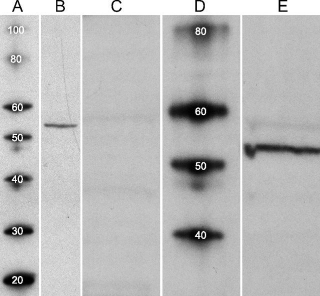
Western blots of D1a dopamine receptor. Homogenate of snap-frozen retinas, and protein standards, separated by SDS-PAGE and transferred to nitrocellulose membranes. A, D, Molecular weight (MW) standard proteins, with MW of each indicated in kilodaltons by superimposed number. B, Retina proteins run alongside standard proteins in A and probed with anti-D1a receptor antibody. A well focused protein band is seen at migration distance corresponding to an estimated MW of 54 kDa. No other proteins are stained over the MW range shown (20–100 kDa). C, E, In a different experiment, retina proteins run alongside standard proteins in D. Lane C probed with anti-D1a receptor antibody that had been preincubated overnight with immunogen. Probing of lane E with anti-D1a receptor antibody shows a well focused protein band in E at an estimated MW of 54 kDa. A faint band is also seen within the MW range reported for glycosylated D1a receptors (here between 55 and 60 kDa). Staining of both bands (dark and faint) was blocked completely by immunogen (C).

