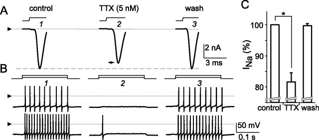
Figure 10.
Reduction of voltage-gated Na+ current and spiking by nanomolar tetrodotoxin. Recording mode, conditions, and figure format are as in Figure 8. A, Voltage-gated Na+ current (without leak subtraction) and spikes elicited in a single ganglion cell by depolarizations in voltage- and current-clamp modes, respectively. Current activated by voltage jump from −72 to −47 mV as solution superfused over the cell is changed from control (A1) to 5 nm TTX (A2) and then control again (A3). The triangle is positioned at zero current level. The dashed horizontal line at peak of control current highlights partial reduction of current amplitude by TTX and full recovery during wash. B, Spikes then elicited in the same cell by constant current injections (10 and 30 pA) as solution superfused over the cell is changed from control (B1) to 5 nm TTX (B2) and control (B3). At this concentration, and as seen during the response to dopamine in other cells, TTX reversibly reduced peak current amplitude by 14%, raised spike threshold (viz., abolished spiking elicited by smallest current injections), and curtailed spiking elicited by larger current injections (bottom trace, middle column). The triangles are positioned at zero voltage level for all traces in each row. C plots mean (solid bar) and SEM (error bar) of peak inward current during microperfusion of control solution, TTX (4–5 nm), and after wash with control solution, for all cells tested (n = 3). The means in control and TTX differed significantly (*p < 0.0001, paired t test).

