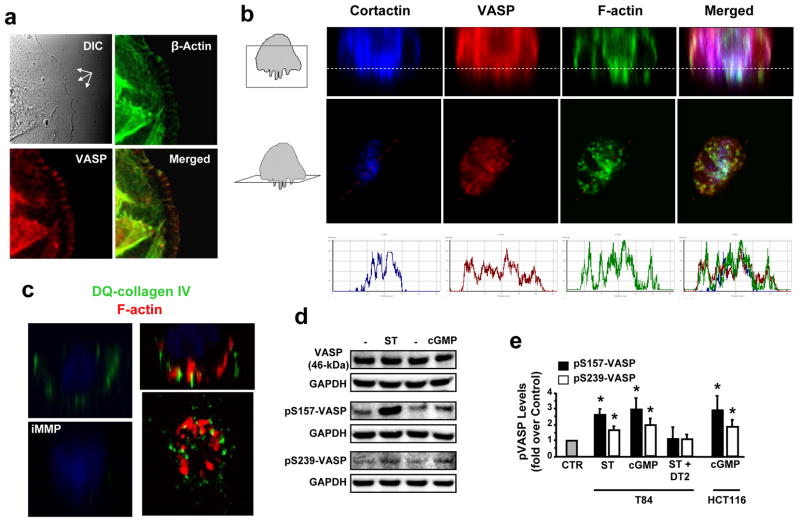
Fig. 1.
VASP Ser phosphorylation is induced by GCC activation in colon cancer cells. (a) Cell leading edges (multi-arrows in upper-left panel) of T84 human colon carcinoma cells (on 2-D surfaces) were imaged (magnification, 63x) by DIC and confocal microscopy following immunofluorescent staining with phalloidin (green) and anti-VASP antibody (red). Yellow in merged image indicates F-actin and VASP colocalization at leading tips. (b) Representative confocal microscopy images of invadopodia from T84 cells grown on 3-D Matrigel scaffolds (Matrigel/air interfaces, dotted lines in top panels). Boxes in cartoons on left indicate visual planes in vertical (top panels) and horizontal (middle panels) cross-sections from Z-stack images. Cells were immunofluorescently stained for invadopodial marker cortactin (ref. 27) (blue), VASP (red) and F-actin (green). In merged images, white (upper panels) and coincidence of picks (bottom panels) from distinct fluorescent signals along lines depicted in middle panels indicate cortactin, VASP and F-actin colocalization. (c) Vertical or horizontal (bottom-right) T84 cell cross-sections of confocal microscopy images obtained as in b. Here, Matrigel contained DQ-collagen IV, which releases a fluorescent product (green) upon cleavage by extracellular proteases. Application of 60 nM of the broad MMP inhibitor GM6001 (iMMP) was employed to prevent DQ-collagen IV degradation by tumor cells (ref. 20) and serve as the negative control condition. Red in right panels reflects F-actin staining. (d) Immunoblots of a representative experiment with T84 cells, treated with the GCC ligand ST (1 μM, 5 min) or the cGMP analog 8-br-cGMP (5 mM, 30 min). pS157-VASP and pS239-VASP, VASP phosphorylated at Ser157 and Ser239, respectively; GAPDH, the loading control. (e) Bar graphs reflect densitometric quantification of immunobands, normalized to respective loading (GAPDH) and vehicle (PBS) controls (CTR), from experiments with T84 and HCT116 colon cancer cells treated as in d. Some conditions also received the peptide DT2 (5 μM, 30 min) to selectively and completely inhibit PKG-Iα activity (Ki, 12.5 nM; ref. 17). *, P < 0.05, versus respective PBS control.

