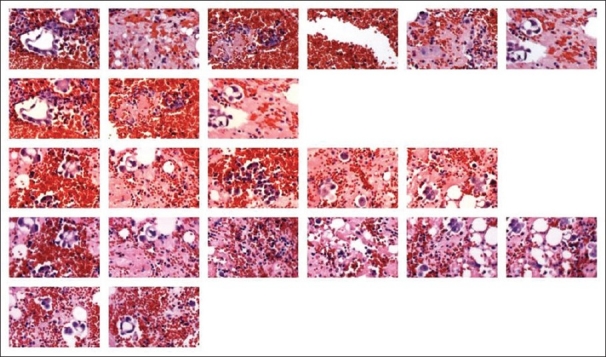
Figure 3.

Malignant pleural effusion of a patient with lung adenocarcinoma. A cell block of a pleural effusion from a patient with metastatic lung adenocarcinoma was stained with H and E. Multiple images representative of different fields of view were taken using the camera on the Arcturus XT™ LCM stage. The slide was not coverslipped and was coated with xylene. The images were then “autocorrected” using Microsoft Picture Manager. dCORE was used to capture representative malignant and benign features, which were then loaded into IMA to create the image array shown here.
