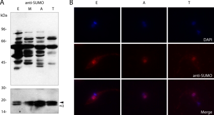
Fig. 2.
Analysis of SUMOylation pattern in life-cycle stages of T. cruzi. A, Total cell lysates of 2 × 10⁷ epimastigotes (E), metacyclic trypomastigotes (M), amastigotes (A), and cell-derived trypomastigotes (T) were electrophoresed on SDS-PAGE 7.5% (upper panel) and 15% (lower panel), transferred to nitrocellulose membranes and probed with anti-TcSUMO polyclonal antibodies. Precursor and mature forms of SUMO are depicted with a filled or empty arrowhead, respectively. B, Indirect immunofluorescence study of the different forms of the parasite using anti-TcSUMO polyclonal antibodies and AlexaFluor 546-conjugated goat anti-mouse secondary antibody. Nuclear and kinetoplast DNA were visualized by DAPI staining. The SUMO fluorescence image (red) has been merged with the corresponding DAPI staining (blue), and the resulting merged images are shown.

