Fig. 3.
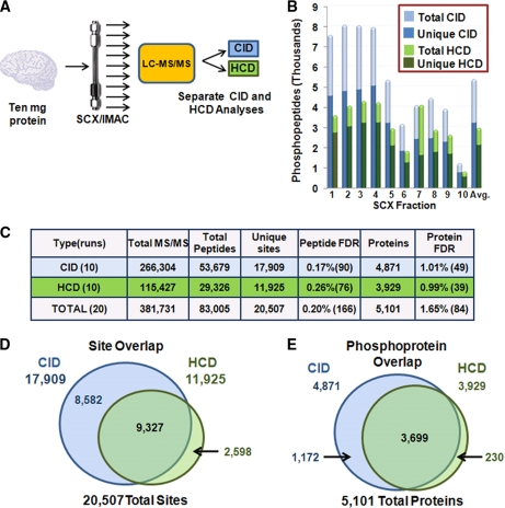
Full phosphoproteome analysis of mouse brain by CID- and HCD-type fragmentation. A, Workflow for phosphoproteomic analysis. Brain phosphopeptides were enriched with the SCX-IMAC approach (26), and ten fractions were analyzed by separate 85-min CID and HCD runs. B, Phosphopeptides identified in SCX fractions. C, Summary of these studies. Matches to reversed (decoy) sequences are in parentheses. D, E, Venn diagrams of site and phosphoprotein overlaps between CID and HCD experiments.

