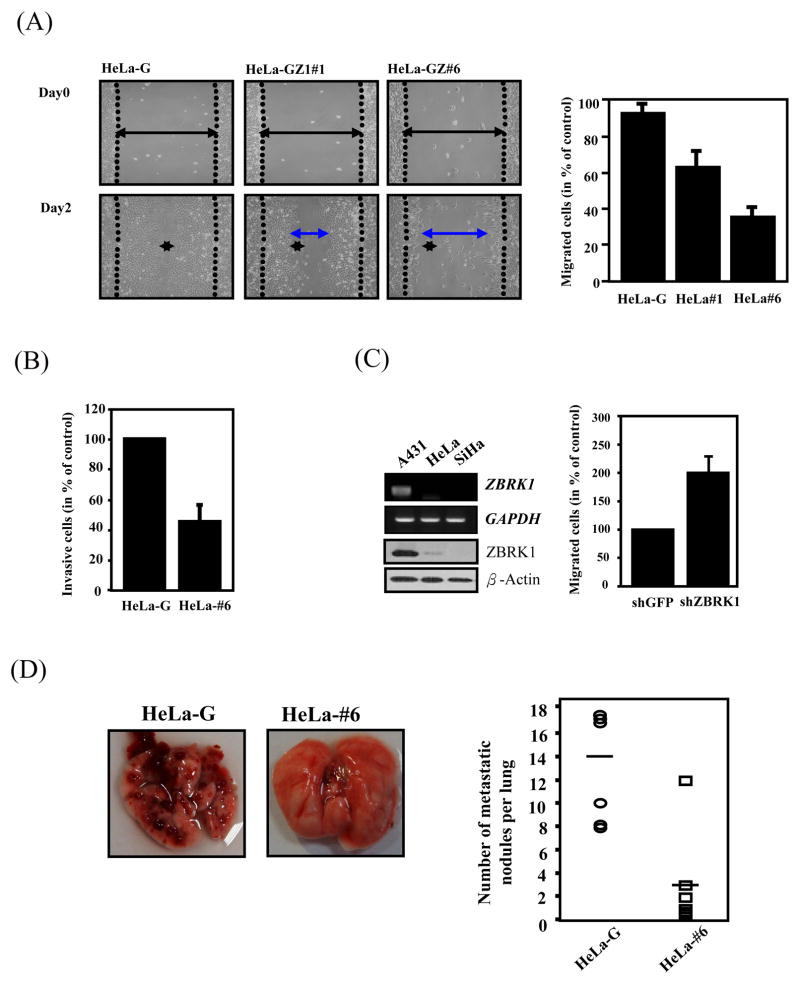
Figure 3. ZBRK1 inhibits cell migration.
(A) Wound-healing migration was performed with EGFP- and EGFP-ZBRK1-expressing cells. Representative images of wound sealing were taken on the day of the laceration and day 2 after the wound scratch. The level of cell migration into the wound scratch was quantified as the percentage of wound sealing. Values represent the average ±S.E.M. of three independent measurements. (B) Overexpression of ZBRK1 inhibits invasion of cancer cells. The cells were seeded in ECMatrix layer and the level of cell invasion was determined using QCM™ 96-well cell invasion assay as described in Materials and Methods. (C) Inactivation of ZBRK1 increased migration of cancer cells. Left panel: mRNA and protein levels of ZBRK1 in A431, HeLa, and SiHa cervical cancer cell lines. Right panel: A431 cells were treated with lentiviral shZBRK1 or shGFP in the QCM™ 96-well cell migration assay. The top panel shows the amounts of ZBRK1 and GAPDH. (D) Equal amounts of EGFP- and EGFP-ZBRK1-#6 HeLa cells were injected into the tail vein of scid mice. The experimental mice were sacrificed to calculate the metastatic nodes on lung tissues after 4 weeks. Data from six mice in each group are presented as the mean ± SD. Representative pictures taken at the time of sacrifice are shown (left panel).

