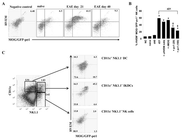
Fig. 7.
HVEM+ IKDCs bind MOG-pσ1. A. Splenic and LN lymphocytes from naïve- or EAE- challenged mice 10 or 40 days earlier were tested for their ability to bind MOG/GFP-pσ1 following 4 days restimulation with MOG35–55 and IL-2. Negative control cells were stained with MOG-pσ1 instead of MOG/GFP-pσ1. Data depict cells gated on NK1.1+ cells (representative of four mice/group). B. Percentages of NK1.1 HVEM+ cells binding to MOG/GFP-pσ1 were assessed. Anti- HVEM mAb (20 μg/ml), recombinant MOG (20 μg/ml), or recombinant pσ1 (20 and 100 μg/ml) was added when indicated. Each column represents the mean ± SD from 4 mice/group. **P ≤ 0.01 versus negative control (NC); ‡P < 0.01 versus d21 MOG/GPF-pσ1 staining. (C) Splenocytes from a mouse (representative of 3 mice) at the peak of the disease were gated on CD11c+ DCs, CD11c+NK1.1+ cells (IKDCs), and NK1.1+ CD11c− NK cells and analyzed for MOG/GFP-pσ1 binding and HVEM expression.

