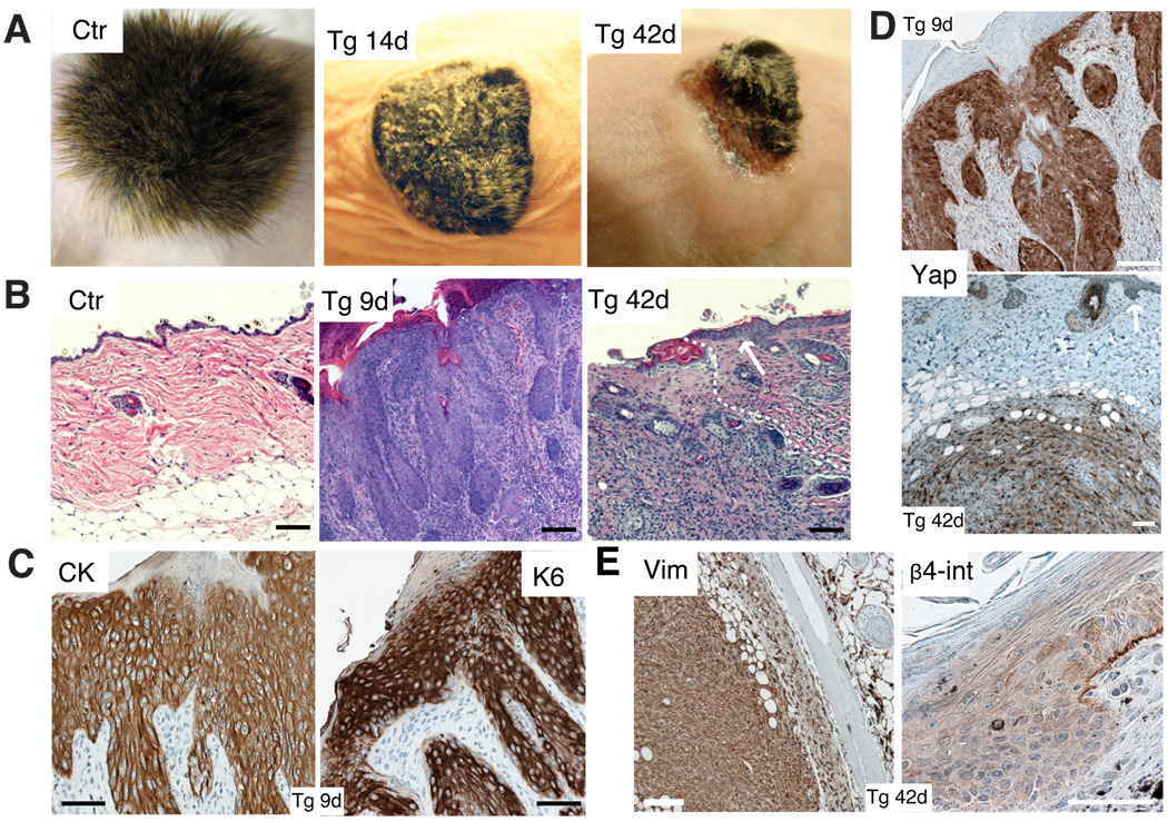
Figure 2. Activation of Yap1 leads to tumor formation.
A, Gross morphology of Ctr and Tg grafts 14 and 42 days after beginning of Dox treatment. Note hyperkeratosis and stunted hair growth in Tg 14d graft and ulceration of skin and subcutaneous tumor mass in Tg 42d graft. B, H&E staining of Ctr and Tg grafts 9 and 42 days after Dox induction show an invasion of underlying dermis by invasive keratinocytes derived from the Tg graft. Arrow points out to neighboring normal Nude mouse epidermis. White dashed line indicates border of carcinoma to normal Nude dermis.C, Expression of cyto-keratin (CK) and keratin 6 (K6) in Tg grafts treated 9 days with Dox as shown by immunohistochemistry. D, Immunohistochemistry for Yap1 on Tg grafts with 9d and 42d Dox treatment reveals epithelial origin of the tumor. Arrow points at Nude mouse epidermis. E, Immunohistochemistry for vimentin (Vim) and integrin-β4 (β4-int) on Tg grafts with 42 days Dox induction. Scale bars represent 100 µm. See also Supplementary Figure S1D–H.

