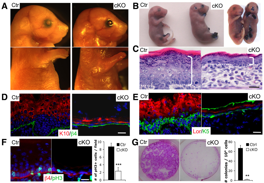
Figure 3. Yap1 is required for the maintenance of the proliferative capacity of epidermal stem cells.
A, Gross morphology of representative control (Ctr) and cKO E18.5 embryos. Note absence of skin in distal limbs, eyes, and ears and overall thinner skin. B, Toluidine blue skin barrier assay in E18.5 mice reveal absence of epidermal barrier in limbs, ears, nose and mouth. C, H&E staining shows a decrease in epidermal thickness in cKO skin compared to the Ctr (indicated by bracket). Note the flat morphology of basal cells and disorganized epidermal architecture. Dashed line represents epidermal-dermal junction. D-E, Immunofluorescence on frozen E18.5 epidermis reveals signs of basal cell depletion in cKO skin. Note the reduction of keratin 10 (K10) negative, keratin 5 (K5) positive cells progenitor/stem cells juxatposed to the basement membrane (β4). Note background staining for K5 in stratum corneum of cKO. F, Marked reduction in number of proliferative basal cells in cKO limb skin as measured with an antibody against phospho-histone H3 (pH3). Results represent measurements of at least 5 different fields in 3 different animals. G, Reduction in the number of colony-forming progenitors in E18.5 skin. Rhodamine B stain and colony counts were performed at day eight after plating and are representative of two independent experiments. Data are presented as mean ± standard deviation (error bars). **p value < 0.01, *** p< 0.005. Scale bars represent 20 µm. See also Supplementary Figure S2A–F.

