Figure 1.

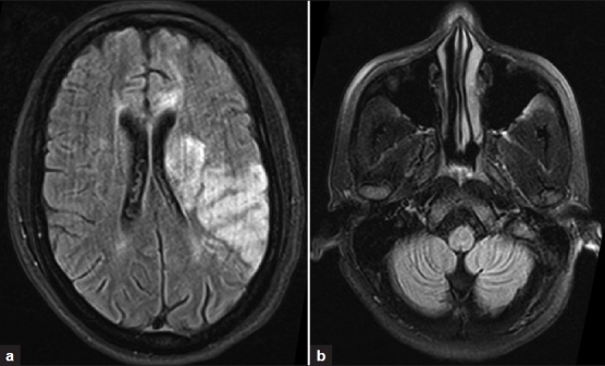
MRI of the brain (a) showing altered signal intensity in the left parietal, temporal cortices and left basal ganglia. Both cerebellar hemispheres (b) are normal

MRI of the brain (a) showing altered signal intensity in the left parietal, temporal cortices and left basal ganglia. Both cerebellar hemispheres (b) are normal