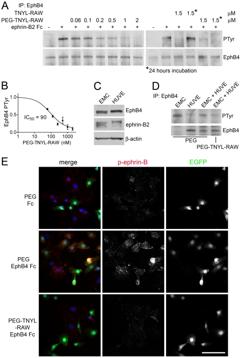
Figure 4. PEGylated TNYL-RAW inhibits tyrosine phosphorylation of EphB4 and ephrin-B2.
(A) B16 melanoma cells pretreated with the indicated concentrations of PEG-TNYL-RAW or TNYL-RAW for 15 min or 24 hours were stimulated with 1.5 µg/ml preclustered ephrin-B2 Fc (+) or Fc as a control (−) for 20 min in the continued presence of the peptide. EphB4 immunoprecipitates were probed with anti-phosphotyrosine antibody (PTyr) and reprobed for EphB4. (B) The inhibition curve shows the relative levels of EphB4 phosphorylation in the presence of different concentrations of PEG-TNYL-RAW, which were quantified from immunoblots and normalized to the amount of immunoprecipitated EphB4. Error bars represent the standard error from 3–6 experiments. (C) HUVEC and EMC lysates were probed for EphB4, ephrin-B2 (band at ∼45 Kd detected with a pan-ephrin-B antibody) and ß-actin as a loading control. It is not known why the ephrin-B2 band appears as a more prominent doublet in EMCs than HUVECs. (D) HUVECs and EMCs were cultured individually or mixed at a 1∶1 ratio in the presence of 1.5 µM PEG-TNYL-RAW or PEG control. EphB4 immunoprecipitates were probed with an anti-phosphotyrosine antibody (PTyr) and reprobed for EphB4. (E) HUVECs and EMCs, which express EGFP, were cultured at a 1∶1 ratio for 15 hours in the presence of 1.5 µM PEG-TNYL-RAW or PEG control. The cells were then stimulated with 1.5 µg/ml preclustered EphB4 Fc or Fc as a control for 20 min in the continued presence of the peptide or PEG. The cells were stained for phospho-ephrin-B (red), which likely corresponds to the phosphorylated form of the EphB4 preferred ligand ephrin-B2, and nuclei were labeled with DAPI (blue). Scale bar = 50 µM. Fluorescence intensity from 6 micrographs per condition was quantified. The values obtained (expressed in arbitrary units ± standard error) are: PEG & Fc, 24±2.6; PEG & EphB4 Fc, 44±3.6; PEG-TNYL-RAW & EphB4 Fc, 20±2.5. The fluorescence of cells treated with PEG-TNYL-RAW & EphB4 Fc was significantly (P<0.001) different from that of cells treated with PEG & EphB4 Fc, but not from that of cells treated with PEG & Fc, by one-way ANOVA and Bonferroni's post test.

