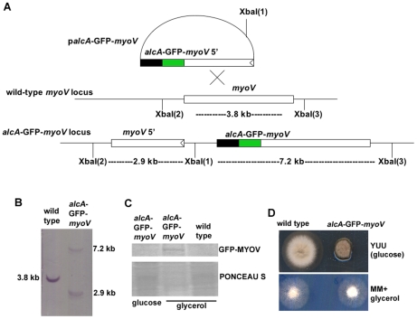
Figure 1. Construction of the alcA-GFP-myoV strain.
(A) A diagram showing the homologous integration of the palcA-GFP-myoV plasmid into the genome (see Materials and Methods for details). (B) A Southern blot confirming the homologous integration event. (C) A Western blot showing that the GFP-MYOV fusion protein can be detected in extracts of cells grown on glycerol but not on glucose. A protein extract from a wild type strain grown on glycerol was use as a negative control for the anti-GFP antibody. PONCEAU S staining of the same blot is shown as a loading control. (D) Growth phenotypes of the alcA-GFP-myoV strain grown on glucose (YUU) and glycerol (MM+glycerol) plates at 37°C for 2 days. The strains were point inoculated on different plates. Note that on YUU, the growth of the mutant is significantly reduced, but on MM+glycerol, the mutant colony is almost identical to a wild type colony.

