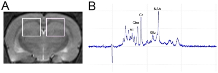
Figure 5. Illustration of the voxel to the site of hippocampus for 1H-MRS was presented, with associated magnetic resonance spectrographic imaging spectrum.
(A) Left and right hippocampi are indicated by the white rectangles in coronal T2-weighted imaging of rat brain. (B) Typical hippocampal 1H-MRS with marked Myo-inositol (MI), choline-containing compounds (Cho), creatine and phosphocreatine (Cr), glutamate (Glu) and N-acetylaspartate (NAA) peaks.

