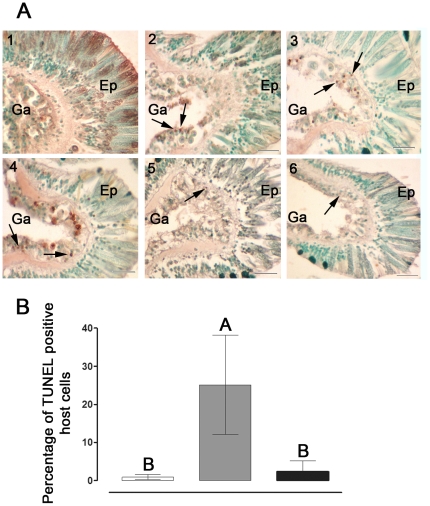
Figure 3. Detection and quantification of apoptosis-like activity in S. pistillata.
(A) Temperature-induced DNA fragmentation detected in situ in tissues of S. pistillata. Fragments were incubated in control (24°C) or thermal stress (34°C). Sections (7 µm-thick) were attached to slides and apoptotic cells were detected using the TUNEL technique. (1) control; (2) 6 h, (3) 24 h, (4) 48 h, (5) 72 h and (6) 168 h of thermal stress. Arrowheads indicate apoptotic nuclei stained brown, while intact nuclei are counterstained green. Scale bar, 20 µm. Ep, epithelium. Ga, Gastroderm. (B) Percentage of TUNEL positive cells in tissues of S. pistillata incubated in control (24°C, white) or subjected to thermal stress of 34°C for 24 h (gray) and 168 h (black). Sections (7 µm-thick) were attached to slides and apoptotic cells were detected using the TUNEL technique. The mean percentage of TUNEL positive cells of individual fragments (n = 3) was derived by comparing the number of TUNEL positive cells to the total number of cells in the same image field. A total of five image fields per fragment section was analysed and averaged. Means were compared using a One-Way Anova with Tukey post-hoc testing. Letters above bars denote statistical significance; two means are significantly different (P<0.05) if their letters are different.

