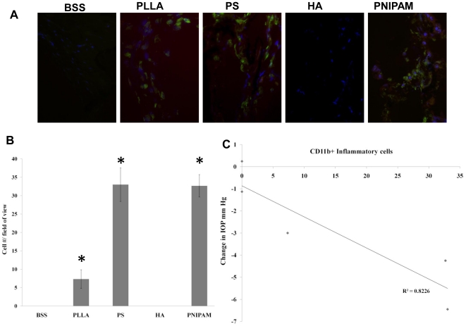
Figure 5. The accumulation of CD11b+ inflammatory cells in the trabecular meshwork.
The representative immunohistochemically stained images showed the accumulation of CD11b+ inflammatory cells (labeled green; DAPI staining to locate cell nucleus) in the trabecular meshwork following particle injection (A). The extent of CD11b+ cell accumulation in the trabecular meshwork was quantified (B). Data are mean ± standard deviation. Significance of PLLA, PS, HA, PNIPAM vs. BSS control; * p<0.05 Correlation between CD11b+ inflammatory cells and average IOP changes in different test groups was also determined (C).

