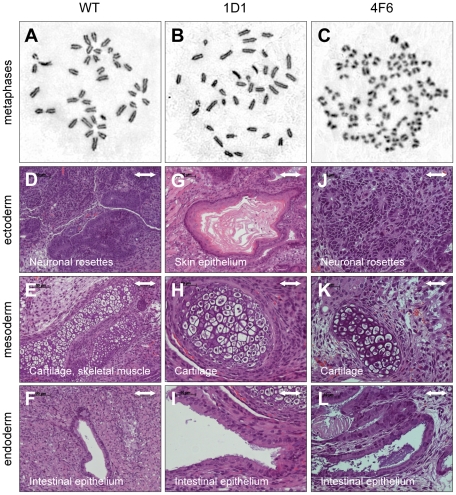
Figure 4. Assessment of metaphase chromosomes and pluripotency.
(A–C) Chromosome analysis. Metaphase spreads of BK4-G3.16 cells were stained with Giemsa. A representative picture of ∼20 analyzed metaphase spreads per clone is shown. (D–L) Assessment of pluripotency. ZFN-treated BK4-G3.16 cells were injected subcutaneously into immunodeficient mice and teratomas removed after 4–8 weeks. Histological analysis using hematoxylin/eosin staining revealed tissues derived from ectoderm (D, G, J), mesoderm (E, H, K) and endoderm (F, I, L). Scale bars = 100 µm for (D–G) and 50 µm for (H–L).

