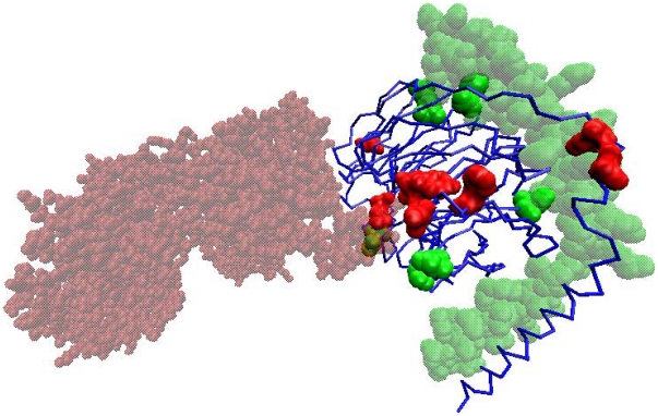
Figure 6. Partner-specific prediction of two interfaces for the beta subunit of the guanine nucleotide binding protein (transducin beta chain 1) (PDB ID: 3PSC, chain B is shown as the blue cartoon).
The two partners are shown in transparent colors (chain A, which is the beta-adrenergic receptor kinase-1, is shown in red, and chain G, which is the guanine nucleotide binding protein's gamma-2 subunit, is shown in green). Predictions from the pair-wise model for each partner chain were converted into single chain predictions and displayed on chain B. Common binding sites, predicted with both partners, were removed and residues exclusively predicted with each partner are shown in the corresponding partner color. Out of the 30 top-scoring residues after removing common predictions, most residues have been assigned to the correct partner.

