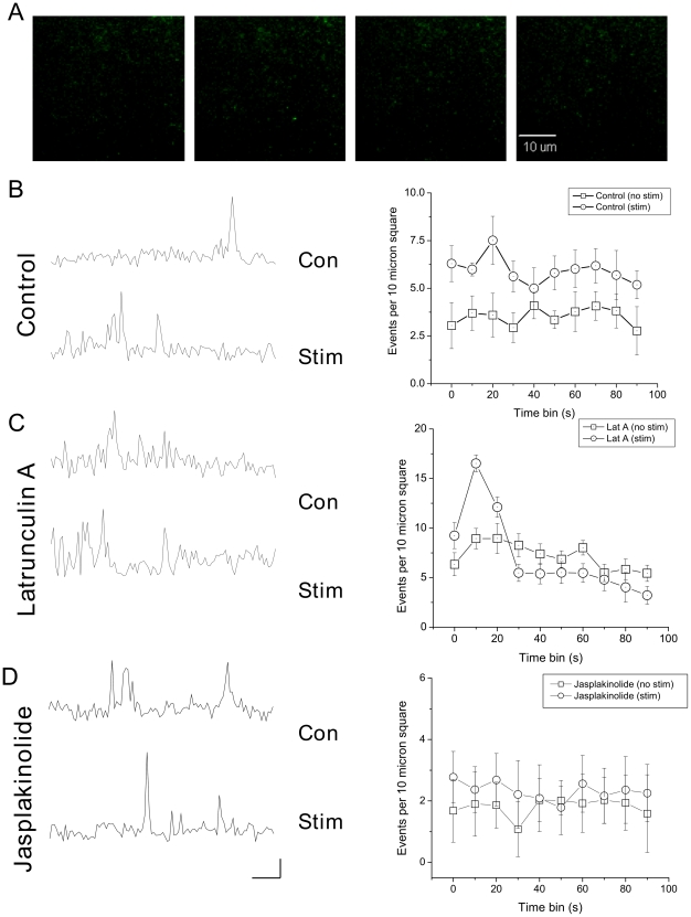
Figure 4. Effect of latrunculin and jasplakinolide on vesicle recruitment and secretion.
(A) Visualized ANF-EmGFP vesicles can be observed arriving and fusing at the plasma membrane. (B) To the left, example ROI traces are shown before and after depolarization with 30 mM K+. To the right, release from 6 cells was analyzed before and after stimulation. Release was normalized as events per 10 µm2, per 10 s. (C) 6 cells were treated with latrunculin A; example ROI traces are shown to the left, and pooled data to the right. Latrunculin treatment caused an increase in spontaneous vesicular secretion; stimulation increased this further, however, this additional release fatigued rapidly (right). (D) 7 cells were treated with Jasplakinolide (10 µM), again, example ROI traces are shown on the left, and time binned averages on the right. Jasplakinolide treatment slightly reduced spontaneous rates of exocytosis, and prevented any significant increase following stimulation. Scale bar shows 10 s/10 arbitrary fluorescence units (directly comparable between conditions).

