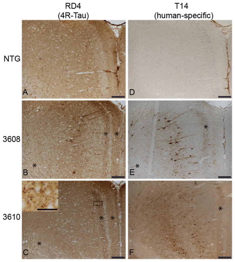
Figure 3.

Somatodendritic distribution of 4R-tau in the cingulate gyrus (CG) of E391 transgenic mice. (A–F) Coronal paraffin sections were stained with the 4R-tau specific antibody RD4, which recognizes both mouse and human 4R-tau (A–C) or with the human specific tau antibody T14 (D–F). Non-transgenic (NTG) mice exhibit a normal axonal distribution of 4R-tau in the CG (A). Somatodendritic distribution of 4R-tau in the CG is detectable with the 4R-tau specific (B) and human tau specific (E) antibodies in E391-3608 mice (lower expressers). Only the human specific tau antibody demonstrates somatodendritic localization in the CG in E391-3610 mice (higher expressers) (C, F). *Granular dot-like neuritic processes in CG and supracallosal regions. Inset in (C) is a higher power of boxed region showing granular dot-like neuritic processes. Scale bars: A–F, 100 μm; inset in C, 25 μm.
