FIGURE 1:
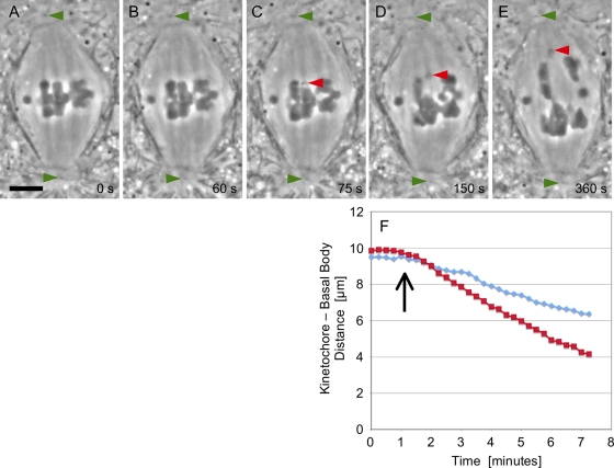
(A–E) Images were taken from a time-lapse movie (Movie S1) of a K fragment generated at anaphase onset. (A) At metaphase, the three autosomal bivalents are positioned at the spindle equator. The middle dichiasmic bivalent is a desirable target for laser cutting, because the chromosome arms are especially thin in the plane just under its kinetochore fiber–attachment sites. (B) At anaphase onset, cohesion between homologues is lost. Measurement of distance between kinetochores and the upper pole is readily achieved using the position of the polar basal bodies as a reference (green arrowhead). (C) Subsequent to the laser flash, the K fragment (red arrowhead) moves poleward, and its severed arms move backward to make contact with its partner homologue. (D and E) Anaphase A continues. The K fragment (red arrowheads) has a poleward velocity twice that of controls. Some collateral damage is evident in the arms of adjacent chromosomes, an unavoidable complication when using the laser beam configured as a line, as it was in this case. Scale bar: 5 μm. (F) Results of frame-by-frame analysis of images yielded these distance vs. time plots for the K fragment (red squares) following detachment (arrow) and for the control kinetochore (blue diamonds) to the left of the fragment in (C).

