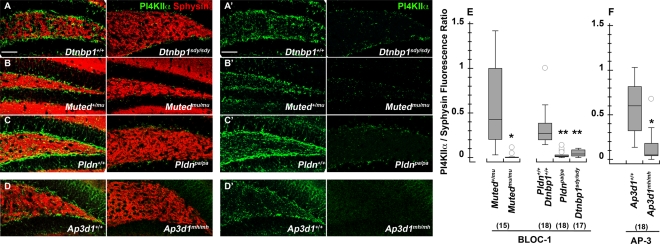
FIGURE 2:
Dentate gyrus PI4KIIα content is reduced in the neuropil of BLOC-1– and AP-3–deficient mice. The dentate gyrus of the hippocampal formation from 6- to 8-wk-old control (A–D), BLOC-1–deficient sandy (Dtnbp1sdy/sdy), muted (Mutedmu/mu), and pallid (Pldnpa/pa) and AP-3–deficient mocha (Ap3d1mh/mh) mice was stained with antibodies against PI4KIIα (green) and the synaptic vesicle marker synaptophysin (red). (E) Total pixels for synaptophysin and PI4KIIα were quantified by MetaMorph analysis and expressed as a ratio of PI4KIIα to synaptophysin pixel counts. Numbers in parentheses represent the number of independent sections stained from three animals. *p < 0.0001; **p < 0.005. Bar, 50 μm.

