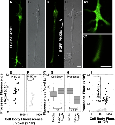
FIGURE 5:
PI4KIIα targeting to neurites in PC12 cells requires the PI4KIIα dileucine-sorting motif. PC12 cells expressing wild-type EGFP-PI4KIIα (A, B; n = 17 cells) or EGFP-PI4KIIαL60-61A (C, D; n = 15 cells) tagged with EGFP were NGF differentiated for 48–72 h posttransfection and cells were imaged in vivo. (B, D) DIC images. (A1, C1) Enlarged view of neurite tips in A and C. (E, F) Fluorescence intensity per voxel was measured for wild-type PI4KIIα-expressing (closed circles) and PI4KIIαL60-61A-expressing (open circles) cells both in cell bodies and their processes. (H,G) Comparison of fluorescence intensity between PI4KIIα- and PI4KIIαL60-61A–expressing cells from E and F for cell bodies and processes, respectively. (I) Transfected cells were stained for VAMP2 and EGFP and imaged by confocal fluorescence microscopy. Fluorescent pixels present in cell body and processes were quantified for both VAMP2 and transfected PI4KIIα. Closed symbols depict data from cells expressing wild-type PI4KIIα (n = 26 cells), whereas open symbols depict fluorescent pixels from cells expressing PI4KIIαL60-61A (n = 26). Circles and triangles represent EGFP and VAMP2 fluorescence values, respectively. In E, F, and I, each point depicts the fluorescence intensity in processes and cell body of individual cells as in terms of x, y coordinates. Data were collected from three independent experiments. Bars, 10 μm.

