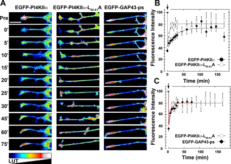
FIGURE 6:
Distinct mechanisms mediate the delivery of wild-type and dileucine mutant PI4KIIα into neurites. (A) Look-up table (LUT) of photobleached neurite tips of PC12 cells expressing EGFP-PI4Kα, EGFP-PI4KIIαL60-61A, or EGP-GAP43-ps. In vivo images were taken before (Pre), during photobleaching (0') and every 5 min thereafter for 30 min, after which an image was acquired every 15 min for an additional 45 min. (B, C) Time course of neurite tip fluorescence intensity (%) during FRAP, normalized to their fluorescence intensity before photobleaching. (B) EGFP-PI4KIIαL60-61A (n = 14 cells) recovers faster than EGFP-PI4Kα (n = 23 cells) following photobleaching, reaching a plateau within 10 min vs. 45 min, respectively. (C) No differences are observed in the time course of recovery between EGFP-PI4KIIαL60-61A and EGP-GAP43-ps (n = 8 cells). Scale bars, 20 μm.

