FIGURE 5:
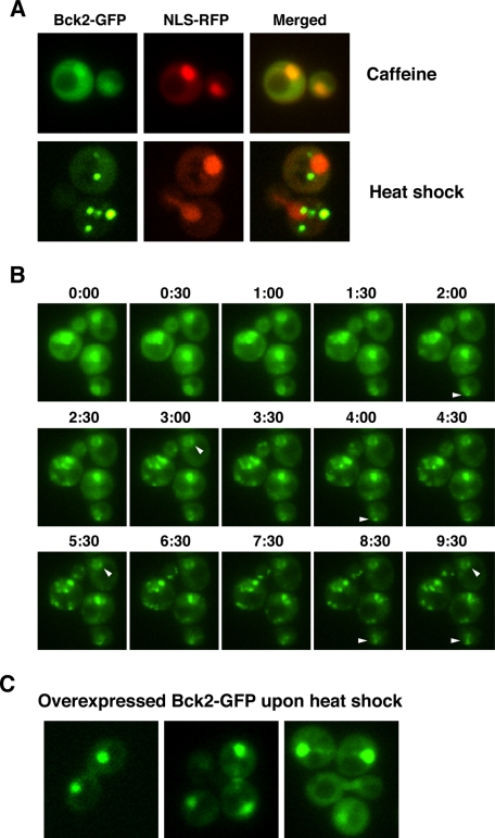
Bck2 localization significantly changes during heat shock. (A) Representative wide-field fluorescence images of Bck2-GFP cells (FLY3503) 90 min after 15 mM caffeine treatment at 22°C and 30 min after cells were transferred to 39°C (heat shock). Caffeine treatment does not induce changes in Bck2 localization, but heat shock causes Bck2 to relocate from the nucleoplasm to bright cytoplasmic puncta. (B) Time-lapse analysis of heat-shocked Bck2-GFP cells. Bck2-GFP cells were heat shocked at T = 0 and images were captured at 30-s intervals. Arrowheads point to representative nuclear foci that remain visible throughout the experiment. These data are also presented in Supplemental Movie S1. (C) Moderately overexpressed Bck2-GFP (from pGPD-BCK2-GFP) does not localize to mitochondria or other cytoplasmic puncta during heat shock (images were captured 15 min after heat stress induction). All images were captured via wide-field fluorescence microscopy. All images in A and C represent single optical sections, and the images in B are merged from 3 × 0.2 μm optical sections.

