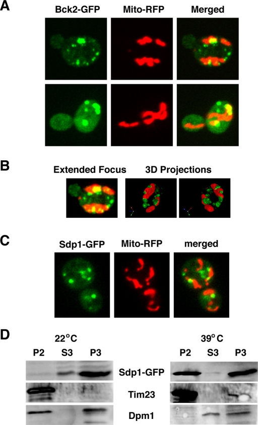
FIGURE 7:
Bck2 and Sdp1 associate with mitochondria during heat shock. (A) Some heat shock–induced Bck2 puncta colocalize with mitochondria. A plasmid expressing an RFP-tagged mitochondrial marker (Mito-RFP; pHCRED) was introduced into Bck2-GFP cells. All images represent single optical sections captured by spinning disk confocal microscopy 15 min after shifting cells to 39°C (heat shock). (B) The cell is presented as a three-dimensional (3D) model via Volocity software (PerkinElmer). Left, a merge/projection of 21 × 0.2 μm Z-sections. Middle and right, different angles of a 3D model of the same cell. See Supplemental Movie S2 for this model in rotation. (C) Sdp1 localizes to mitochondria in heat-shocked cells. Cells expressing Sdp1-GFP and Mito-RFP (FLY3570, plasmids pRS425-SDP1-GFP, pHCRED) were monitored after 15 min of heat stress, as described in A. (D) Immunoblots of organelle fractions of unstressed (22°C) and heat-shocked (39°C) Sdp1-GFP cells (see Materials and Methods). Western blots are probed with antibodies to GFP, Tim23 (a mitochondrial marker), and Dpm1 (ER/microsome marker). Mitochondria are enriched in P2, microsomes and ER are enriched in P3, and S3 is enriched for cytosolic proteins. Note that Sdp1 is enriched in fraction P2 (mitochondria) of heat-shocked cells and not in P2 of unstressed cells. Some Sdp1 is also present in fraction P3 (ER/microsome), regardless of heat shock.

