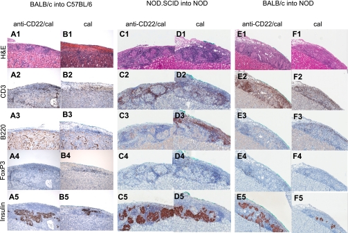
FIG. 3.
Islet pathology in the three immune settings of islet transplantation (allo, auto, and allo/auto). Islets from BALB/c or NOD.SCID mice were transplanted under the kidney capsule of chemically induced hyperglycemic C57BL/6 or spontaneously hyperglycemic NOD mice treated with anti-CD22/cal or cal and were retrieved for histological analysis at different time points (n = 3 mice/group). Grafts from both BALB/c into C57BL/6– and NOD.SCID into NOD–transplanted mice were collected 14 days after transplantation, whereas BALB/c into NOD grafts were collected at day 7 at the time of graft rejection of control mice. 1) In allo at day 14, anti-CD22/cal–treated mice displayed preserved islet architecture (A1) with mild CD3+ cell infiltrate (A2), no B220+ cells (A3), and few FoxP3+ cells (A4). Insulin staining was clearly preserved (A5). Cal-treated and untreated mice showed severe cellular infiltration with a complete disruption of islet architecture (B1) and several CD3+ (B2) and B220+ cells (B3); some CD3+ cells also coexpressed FoxP3+ (B4). Very few cells were positive for insulin (B5). 2) In auto at day 14, anti-CD22/cal–treated mice displayed clear preservation of islet structure (C1) with several CD3+ cells in the infiltrate (C2) surrounding the islets, along with few B220+ (C3) and FoxP3+ cells (C4) as well as abundant insulin-positive cells (C5). Cal-treated mice maintained preservation of islet structure (D1) with insulin-positive cells (D5) but with a massive CD3+ (D2) and B220+ (D3) cell infiltrate and with some FoxP3+ (D4) cells evident in the islet mass. 3) In allo/auto at day 7, islet architecture appeared preserved in anti-CD22/cal–treated mice (E1) with clear insulin staining (E5) but with severe CD3+ cell (E2) infiltrate and few FoxP3+ cells (E4) surrounding the islets, whereas B cells were absent (E3). Conversely, cal-treated mice displayed equally distributed CD3+ (F2) and B220+ (F3) cell infiltration, resulting in complete disruption of islet architecture (F1) and scant insulin staining (F5). Almost no infiltrating FoxP3+ cells were evident (F4). H&E, hematoxylin-eosin. (A high-quality digital representation of this figure is available in the online issue.)

