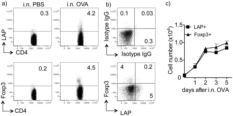
Figure 1. Inhalation of soluble antigen induces LAP+Foxp3− CD4+ T cells.
Naive CD4+CD25− T cells isolated from Thy1.2 OT-II TCR transgenic Foxp3/GFP reporter mice were transferred into Thy1.1 recipient mice. Recipient mice were then tolerized by exposure to soluble OVA (100 μg) in PBS given i.n. for 3 consecutive days. Control (non-tolerized) mice were exposed to PBS without OVA. (a) Representative flow dot plot of LAP and Foxp3 (GFP) expression on gated Thy1.2+ OT-II CD4+ T cells in lung-draining LN from an individual mouse at day 5 after the initial exposure to OVA. (b) Representative flow dot plot of LAP and Foxp3 (GFP) co-staining (bottom), isotype IgG (top) on gated Thy1.2+ OT-II CD4+ T cells in pooled lung-draining LN from an individual mouse at day 5 after the initial exposure to OVA. (c) Total numbers of LAP+Foxp3− and Foxp3+LAP− OT-II CD4+ T cells in lung draining LN populations on different days after the initial exposure to OVA. Data are mean numbers ± SEM from 4 individual mice per group. Data are representative of 3 independent experiments.

