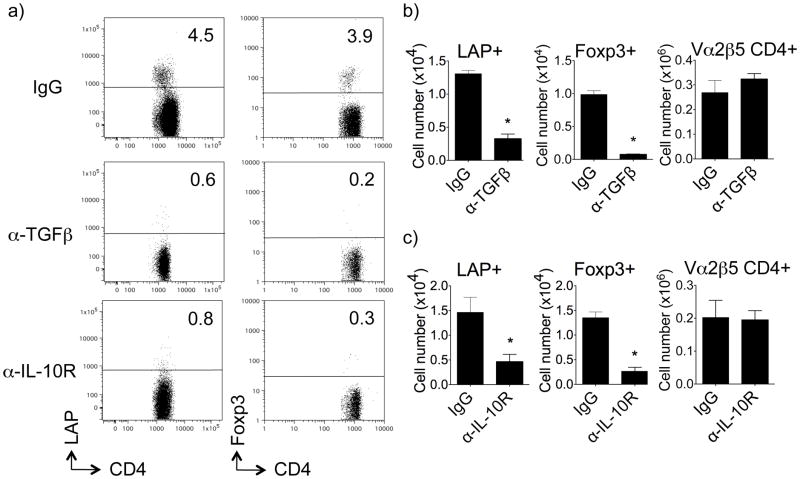
Figure 2. TGF-β and IL-10 are required for induction of LAP+ T cells in vivo.
Thy1.2 OT-II LAP+ and Foxp3+ T cells were tracked in vivo as described in Fig. 1. A single dose of anti-IL-10R (200 μg), anti-TGF-β (200 μg), or control IgG, was given i.p. at the time of initial exposure to i.n. OVA. (a) Representative flow cytometry dot plots of LAP and Foxp3 (GFP) expression on gated Thy1.2+ OT-II CD4+ T cells in lung-draining LN from individual mice. (b) Numbers of LAP+Foxp3− and Foxp3+LAP− OT-II CD4+ T cells, and total OT-II CD4+ cells, were calculated in lung draining lymph nodes on day 5. All results are means ± SEM from 4 individual mice per group. Data are representative of 3 independent experiments. *P < 0.05.

