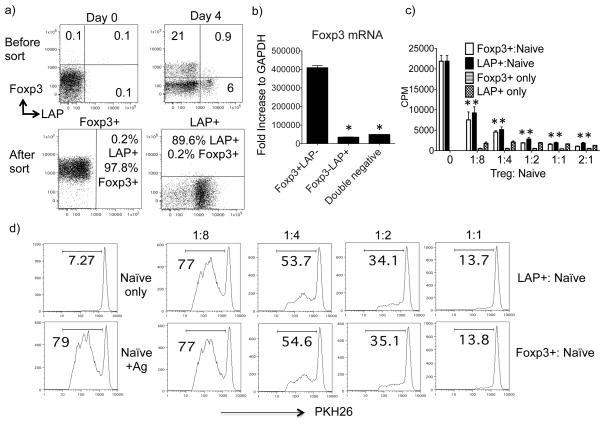
Figure 3. LAP+Foxp3− T cells inhibit naïve CD4+ T cell proliferation in vitro.
Naïve CD4+ T cells from OT-II TCR transgenic GFP/Foxp3 reporter mice were cultured with T-depleted splenocytes and OVA peptide (1 μM) in the presence of exogenous TGF-β (10 ng/ml). Cells were analyzed and sorted on day 4. (a) Top: Foxp3 versus LAP staining on CD4 T cells before and after culture. Bottom: Purity of the sorted LAP+Foxp3− and LAP−Foxp3+ CD4+ T cell populations. (b) Foxp3 mRNA levels in sorted LAP+Foxp3− and LAP−Foxp3+ populations, analyzed using real-time PCR. Data are normalized to the housekeeping gene GAPDH. (c–d) FACS sorted LAP+Foxp3− and LAP−Foxp3+ OT-II T cells (1 × 10⁵) were cultured, alone or at varying ratios with naïve CD4+CD25− OT-II T cells, plus irradiated (3000 rads) syngeneic splenocytes in the presence of 0.5 μM OVA peptide. (c) Proliferation was assessed after a pulse with [3H] thymidine for the last 16 h of a 72-h incubation period. Data are presented as means ± SEM. (d) Responder naïve T cells were labeled with PKH26 dye and division was assessed by loss of the dye after 3 days. Data represent one out of three independent experiments. *P < 0.05.

