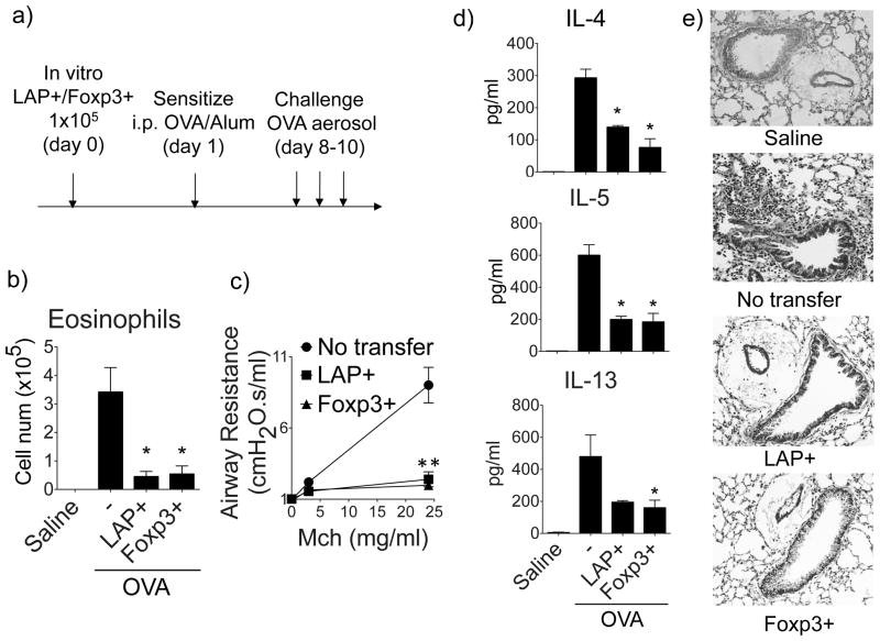
Figure 5. LAP+ T cells prevent induction of asthmatic lung inflammation in vivo.
LAP+Foxp3− or Foxp3+LAP− OT-II T cells, generated in vitro as described in Fig. 3, were sorted and transferred into naïve BL/6 recipient mice. One day later, the mice were then sensitized with OVA (20 μg) in Alum (4 mg) and subsequently challenged with OVA aerosol to induce lung inflammation. (a) Protocol timeline. (b) Eosinophils in BAL. (c) Airway hyperresponsiveness to Methacholine, assessed using a FlexiVent. (d) Cytokines in BAL by ELISA. (e) Representative H&E staining of lung sections. Data are means ± SEM from 3 mice per group. Similar data were generated from 3 independent experiments. *P < 0.05.

