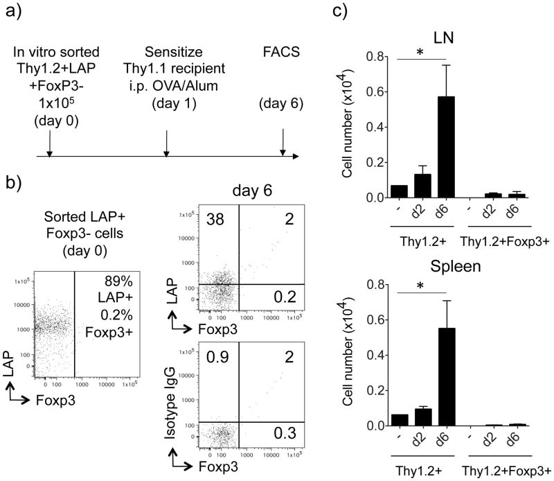
Figure 6. LAP+ T cells do not convert to Foxp3+ T cells in vivo.
Sorted Thy1.2+LAP+Foxp3− T cells, generated in vitro as described in Figs. 3 and 5, were transferred into Thy1.1 recipient mice. One day later, the recipient mice were immunized with OVA (20 μg) and alum (4 mg). (a) Protocol timeline. (b) The expression of surface LAP and intracellular Foxp3 on gated Thy1.2+ T cells was analyzed in pooled LN and spleen on day 6 after the immunization. Representative dot plot of T cells before and 6 days after the transfer. Isotype control staining for LAP shown in bottom plot. (c) Total numbers of recovered Thy1.2+ T cells in pooled lymph nodes and spleens on day 2 and day 6, compared to recovered Thy1.2+ Foxp3+ T cells. All results are means ± SEM from 4 individual mice per group. Data are representative of 3 independent experiments. *P < 0.05.

