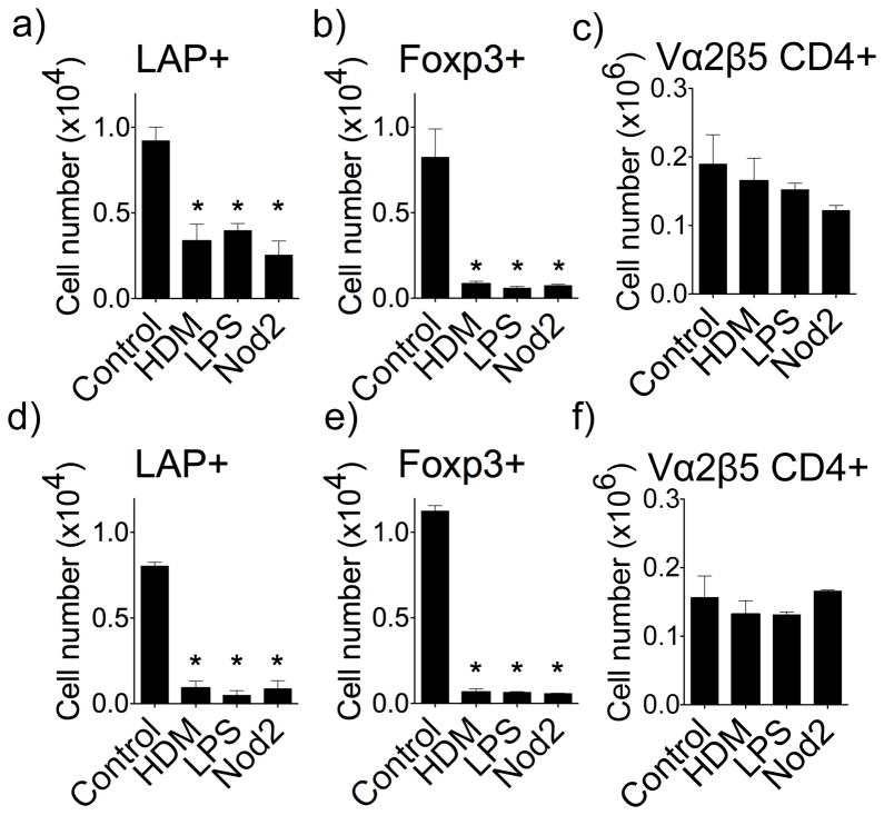
Figure 8. Allergens and pro-inflammatory microbial-associated molecular patterns suppress the generation of LAP+ Treg cells.
The generation of LAP+Foxp3− and Foxp3+LAP− OT-II T cells was tracked in vivo as described in Fig. 1. HDM extract, MDP, or LPS, were given i.n. concurrently with soluble OVA. (a–f) Numbers of LAP+Foxp3−, LAP−Foxp3+, and total OT-II CD4+ T cells in lung draining lymph nodes (a–c) and lung tissue (d–f). Data are means ± SEM from 3 mice per group. Data are representative of 3 independent experiments. *P < 0.05.

