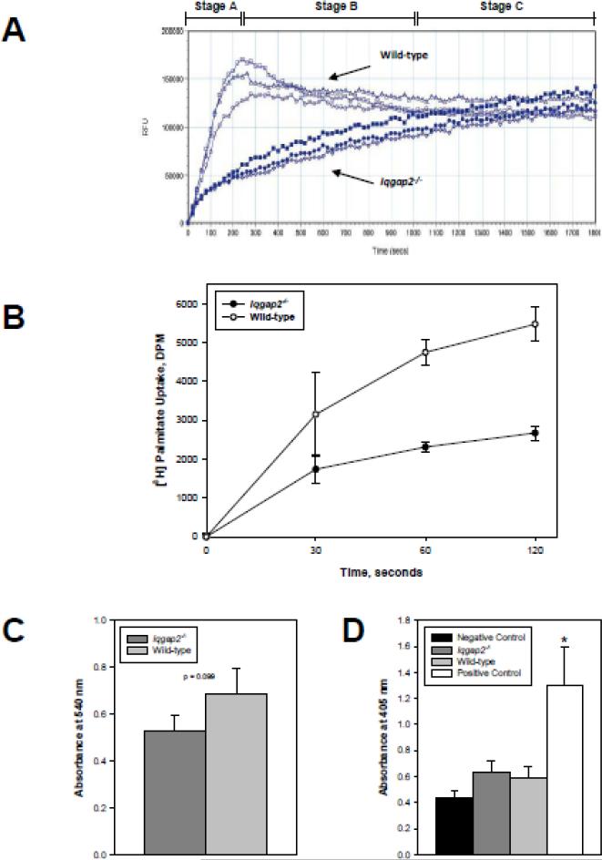
Figure 2.
Selective loss of facilitated fatty acid uptake in Iqgap2-/- hepatocytes. (A) LCFA uptake by isolated hepatocytes from Iqgap2-/- and wild-type mice was measured in real time using BODIPY-FA/Q-Red.1 [32]. Data plots reflect those of a complete set of paired experiments representative of six identical experiments. All data points are mean relative fluorescence units (RFU) per 105 cells/well from triplicate wells (SEM not shown to minimize plot complexity). Three empirical phases of uptake (A, B, C) are shown.
(B) [3H]palmitate uptake by Iqgap2-/- and wild-type hepatocytes. Uptake is expressed as mean decay per minute (DPM) per 2 × 10⁵ cells ± SEM from triplicate wells. p < 0.05 for 60 and 120 seconds time points. Plot is representative of four independent experiments.
(C) Viability of isolated hepatocytes was assessed by MTT assay. Data are expressed as mean relative absorbance at 540 nm per 2 x 10⁵ cells ± SEM from triplicate wells. Difference in absorbance between Iqgap2-/- and wild-type is not statistically significant (p = 0.099).
(D) Apoptosis in isolated hepatocytes was quantified by ApoStrand ELISA and expressed as mean relative absorbance at 405 nm per 104 cells ± SEM from triplicate wells. Asterisk indicates p < 0.05 compared with all other groups.

