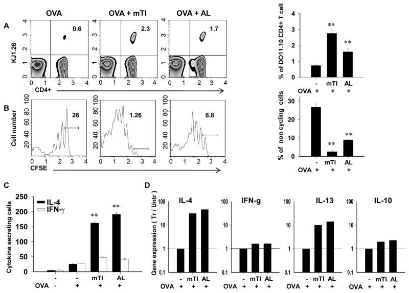
Figure 2. In vivo Ag-specific T cell expansion, cell cycle progression, and IL-4 expression are enhanced by mTi administration.
CD4+ T cells were purified from DO11.10 OVA specific TCR-transgenic mice and labeled with CFSE before transfer to BALB/c recipient mice. Two days later recipient mice were inoculated ip with OVA, OVA + mTi, or OVA + Alum. At day 7 after inoculation, draining mesenteric lymph nodes were removed and stained with anti-CD4-PerCP and KJ-126PE for subsequent analysis. A) T cell expansion was assessed in individual mice within each treatment group and a representative example and the mean and s.e. for four mice/treatment group is shown. B) Cell cycle progression was assessed through decreased CFSE fluorescence and the percent of noncycling cells shown for a representative example and the mean and s.e. for four mice/treatment group. C) MLN cell suspensions from individual mice from each treatment group were cultured with OVA to assess OVA-specific IL-4 and IFN-γ cells per million total cells in each treatment group; mean and s.e. for four mice/treatment group are shown. D) Cytokine gene expression was determined from sorted CD4+, KJ1-26+ MLN T cells (pooled from 5 mice/treatment group) administered OVA, OVA + Ti or OVA + Alum. All experiments were repeated twice with similar results (**p<0.01)

