
Fig. 5.
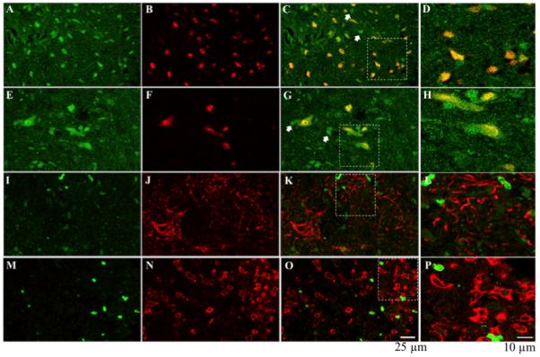
TrkB immunolabeling: TrkB (green; A, E, I & M) is expressed in both dorsal (NeuN: red; B & C) and ventral (red; F & G) neurons, but is not expressed in astrocytes (GFAP: red; J & K) or microglia (OX42: red N & O). Not all TrkB positive labeling is neuronal (white arrows, C & G). To further illustrate TrkB co-labeling with NeuN in the dorsal and ventral spinal cord, GFAP and OX42, the areas indicated by the white rectangles are enlarged in D, H, L & P, respectively.