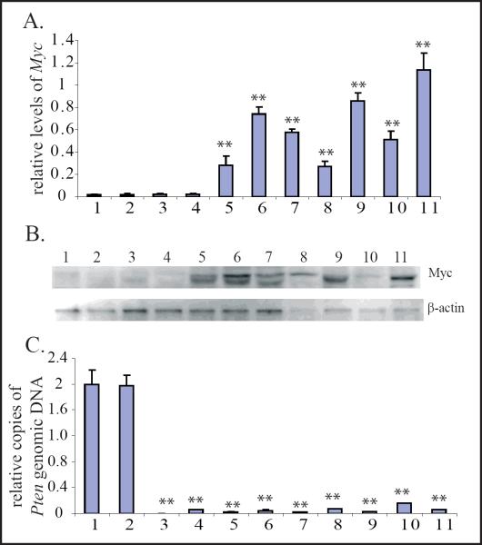
Figure 3.
Myc is up-regulated in leukemic and lymphomic blasts but not in cells from MPDs nor LPDs in Pten−/− mice.A & B. Myc mRNA and protein levels were examined in Gr1+ and CD4+ cells isolated from WT, Pten−/− MPD/lymphadenopathy mice, as well as leukemic/lymphomic blasts from Pten−/− mice using qRT-PCR (A) and Western Blot assays (B.) Relative Myc mRNA levels for each sample were normalized to the mRNA levels of its GAPDH gene. Data are a summary of triplicate experiments (A.) C.Pten deletion in Pten−/− cells was confirmed by q-PCR to detect the levels of Pten genomic DNA. Pten DNA levels for each sample were normalized to GAPDH DNA levels first and then normalized to Pten DNA levels of WT Gr1+ granulocytes (Sample 1.) Samples shown in the figure were: 1. Gr1+ granulocytes from BM of WT controls; 2. CD4+ lymphocytes from lymph nodes of WT controls; 3. Gr1+ cells from BM of Pten−/− MPD mice; 4. CD4+ lymphocytes from enlarged lymph nodes of Pten−/−lymphadenopathic mice; 5–7. CD4+ blasts from enlarged lymph nodes of Pten−/− lymphoma mice; 8. Gr1+ leukemic blasts from BM of Pten−/− AML mice; 9–11. CD4+ leukemic blasts from BM of Pten−/− T-ALL mice. ** indicates statistical significance compared to Gr1+ granulocytes from BM of WT controls.

