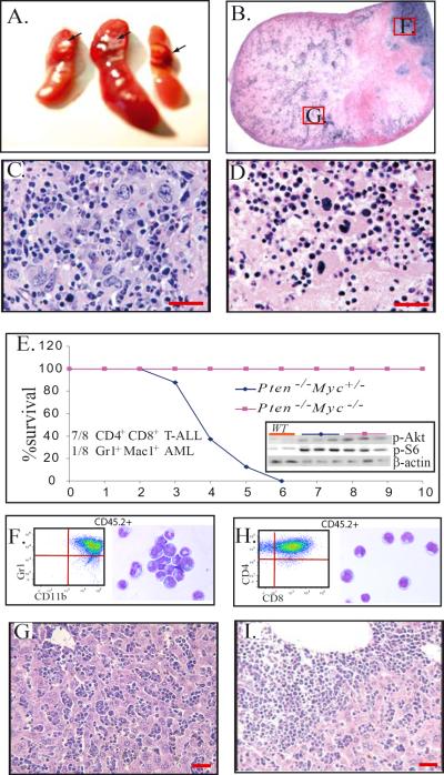
Figure 6.
Myc is required for acute leukemic transformation of Pten-mutant MPDs. A–D. BM NCs from Pten−/−/Myc−/− mice were transplanted into lethally-irradiated mice. Each recipient mouse received 5×10⁶ mutant BM MNCs without support BM cells. Spleens were collected 1 month after transplantation and analyzed by histologic sections. Node-like growths were observed in the recipient spleens (A arrow and B.) Mks were the major component cells in these nodes (C) and the majority of cells in the middle area of the nodes were undergoing apoptosis (D.) E–I. BM MNCs from Pten−/−/Myc−/+ or Pten−/−/Myc−/− mice (CD45.2+) were transplanted into lethally-irradiated recipient mice (CD45.1+) to examine the leukemic transformation of the mutant cells. Each recipient mouse received 5×10⁶ mutant BM MNCs with the support of 2×10⁵ WT BM cells (CD45.1+). Eight mice were transplanted in each group. Recipient mice were monitored for survival (E) and leukemia development. Insert in Fig. E shows increased p-Akt and p-S6 levels in BM cells from both Pten−/−/Myc−/+ and Pten−/−/Myc−/− mice (3 samples from each) compared to WT controls. F–G. Leukemic blasts from BM of Pten−/−/Myc+/− AML (F) and T-ALL (G) mice were analyzed by flow cytometry by gating on the CD45.2+ donor cells. For morphology studies, CD45.2+ donor derived cells were sorted for cytospins. Representative cytospins of blasts isolated from BM of Pten−/−/Myc+/− AML (F) or T-ALL (G) mice are shown by Wright-Giemsa staining. Figure H–I is a representative liver section from a Pten−/−/Myc+/− AML (H) or T-ALL (I) mouse showing leukemic blast infiltration. Bars equal 100 μm.

